M · E · S · M

Minimally Invasive Electro Septal Myectomy

Redefining HOCM surgery
through a single hidden 5cm incision

The world's largest single-center series of minimally invasive septal myectomy for hypertrophic cardiomyopathy, via right infra-axillary incision with transaortic approach.

Zhejiang Provincial People's Hospital, Hangzhou, China

5,000+
MICS procedures
1,000+
HCM myectomies
95%
OR extubation

A right axillary incision

A right axillary incision.
Without sternotomy.

A single 5cm incision is located in the right axilla. The scar is positioned beneath the arm and may be less conspicuous when the arm is at rest. Its appearance and visibility vary with healing, arm position and clothing.

Single
incision
3rd
intercostal space
5
cm hidden incision
Right infra-axillary incision location — hidden beneath the arm

Why MESM

Three core
advantages

MESM bridges the traditional Morrow procedure with the advancements of minimally invasive surgery — offering a unique combination of visualization, instrumentation, and comprehensive repair capability.

1
Superior visualization
Straight-line geometric advantage
The right axillary route provides an endoscopic view through the aortic valve into the left ventricle, supporting assessment of the septum and relevant mitral and subvalvular structures.
2
Purpose-built instrumentation
Sculpts rather than cuts
Electrocautery is used to sculpt the hypertrophied septal muscle.
3
Comprehensive LV repair
Beyond septal reduction alone
Direct visualization enables assessment and treatment of all contributing structures in a single view — septum, abnormal connections and muscle bundles, papillary muscles, mitral leaflets, and chordae. The transaortic approach avoids the long-term uncertainties on mitral valve function associated with a trans-mitral route.

Comprehensive LVOT management

Restore a nearly normal intra-LV anatomy

MESM bridges the traditional Morrow procedure with the advancements of minimally invasive surgery. It addresses the full spectrum of obstructive HCM — including patients with mild-to-moderate septal hypertrophy but severe outflow tract obstruction driven by mitral and subvalvular anatomy. All procedures are performed through the same right axillary approach, in a single operation.

See better, work better.

Septal myectomy
The hypertrophied ventricular septal muscle is sculpted by electro-excision.
Mitral and subvalvular intervention
Anterior leaflet plication or shortening for elongated AMVL. Papillary muscle realignment for anteriorly displaced papillary muscles. Restrictive secondary chordae release when limiting leaflet motion.
Concomitant atrial fibrillation
Surgical ablation, including a Maze procedure, may be considered through the same access in selected patients. Anticoagulation decisions require individual assessment by the treating team; undergoing ablation does not by itself establish that anticoagulation can be stopped.
Multi-valve and combined procedures
Concurrent aortic valve replacement, tricuspid repair, double-valve procedures, ASD closure, and cardiac tumor excision — all via the same minimally invasive access without sternotomy.
Intraoperative checklist — every structure assessed
✓
Septal thickness
✓
Abnormal connections & muscle bundles
✓
Papillary muscles
✓
Mitral leaflets & chordae
Septal myectomy
→
TEE re-evaluation
→
SAM persists?
→
Targeted mitral / subvalvular repair
Live endoscopic view of the left ventricle
Click to play intraoperative video (surgical content)

Endoscopic visualization through the right axillary approach provides an unprecedented direct view of all left ventricular structures — septum, papillary muscles, chordae, and mitral leaflets — enabling comprehensive assessment and repair in a single view. This perspective is unique to the MESM approach and has not been previously described.

Illustrative case

Before and after surgery

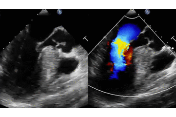
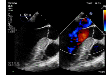
Paired transesophageal echocardiography recordings from one illustrative case.

Preoperative TEE

First frame of the preoperative TEE recording
Still frame
Open original GIF

Postoperative TEE

First frame of the postoperative TEE recording
Still frame
Open original GIF

Clips play independently at their original speed; cardiac cycles are not synchronized.

Clinical outcomes

Evidence

Peer-reviewed publications, international conference presentations, and outcomes from the world's largest single-center minimally invasive HCM surgery program.

Read the RF11 cohort results and their context · Full publication references

LVMCO series — 132 consecutive patients
85→8.5mmHg
Peak gradient
0%
Iatrogenic VSD
62.6min
Mean clamp time
100%
SAM elimination
Key publications
Journal of Cardiac Surgery·2026(1)
Why Septal Thickness Does Not Tell the Whole Story: A Surgeon’s Reappraisal of Dynamic Outflow Obstruction in Hypertrophic Cardiomyopathy
Yong Cui, Zhenzhen Wang, Meijuan Yan
Article 3162395 · Published online November 1, 2026
Narrative review
DOI: 10.1155/jocs/3162395
Annals of Thoracic Surgery·2025
Minimally invasive electro septal myectomy for HOCM through a right infra-axillary incision
100 consecutive cases — presented at STS 2025
Read publication DOI: 10.1016/j.athoracsur.2025.09.023
JTCVS Techniques·2024
An innovative minimally invasive approach for HOCM: transaortic septal myectomy via right infra-axillary incision
Cited by EJHF expert consensus 2026
Read publication DOI: 10.1016/j.xjtc.2024.09.006
Journal of Cardiac Surgery·2022
Minimally invasive right infra-axillary thoracotomy for transaortic septal myectomy
First description of the approach
Read publication DOI: 10.1111/jocs.16546
JASE·2024
Mitral geometry on the mechanism of LVOT obstruction in HCM
Editorial by Sherrid MV
Read publication DOI: 10.1016/j.echo.2024.05.002
JASE·2026
Midsystolic Doppler signal void identifies mid-apical obstruction in HCM
Editorial by Massera & Sherrid
Read publication DOI: 10.1016/j.echo.2026.03.007
EJHF·2026
Expert consensus on diagnosis and management of hypertrophic cardiomyopathy
MESM cited as emerging technique (ref 88)
Read publication DOI: 10.1093/ejhf/xuaf008
International conference presentations
2024
A novel minimally invasive approach for HOCM: transaortic modified Morrow procedure via right infra-axillary incision
AATS 104th Annual Meeting · Toronto
2024
Minimally invasive electro septal myectomy for HOCM via right infra-axillary incision
ISMICS Symposium · Osaka
2024
MESM for hypertrophic obstructive cardiomyopathy
AHA Scientific Sessions · Philadelphia
2025
Minimally invasive electro septal myectomy for HOCM via right infra-axillary incision
STS 61st Annual Meeting · Los Angeles
★ Selected as Best Abstract in Adult Cardiac Surgery
2025
MESM for hypertrophic obstructive cardiomyopathy
ASCVTS Annual Meeting
2025
MESM for HOCM via right infra-axillary incision — China Innovation Session
ISMICS 25th Annual Meeting · Istanbul
2025
Minimally invasive electro septal myectomy for HOCM: 331 consecutive cases
EACTS 39th Annual Meeting · Copenhagen
2025
Minimally invasive electro septal myectomy outcomes
ESC Congress
2026
MESM via right infra-axillary incision: a safe and effective treatment for left ventricular mid-cavity obstruction
AATS 106th Annual Meeting · Chicago · May 3, 2026 · RF11 official abstract
2026
Efficacy and safety of MESM for HCM with residual obstruction after prior septal reduction therapy
AATS 106th Annual Meeting · Chicago
2026
ERAS and ultra-fast-track cardiac anesthesia in LVOTO surgery: 671 patients
ISMICS Annual Meeting

The complete MESM ecosystem

Three disciplines, one team

Surgery, echocardiographic diagnosis, and perioperative care — integrated into a single, seamless pathway from evaluation to recovery.

Dr. Cui Yong
Cui Yong, MD, PhD
Cardiac surgeon
Fellowship-trained at the Mazankowski Alberta Heart Institute, Canada. 5,000+ minimally invasive cardiac procedures, 1,000+ HCM myectomies. Creator of the MESM technique. AATS oral presenter. Member of the Canadian Society of Cardiac Surgeons (CSCS), The Society of Thoracic Surgeons (STS), and the International Society for Minimally Invasive Cardiothoracic Surgery (ISMICS).
Dr. Wang Zhenzhen
Wang Zhenzhen, MD
Echocardiography / Diagnosis
Lead echocardiographer for the HCM program, with work published in the Journal of the American Society of Echocardiography (JASE). Her research focuses on the imaging mechanisms of dynamic outflow obstruction, forming the diagnostic front end of a complete diagnosis-to-treatment pathway for HCM.
Dr. Yan Meijuan
Yan Meijuan, MD
Anesthesia / Ultra-fast-track
Architect of our Ultra-Fast-Track Cardiac Anesthesia (UFTCA) protocol. Over 95% of HCM patients are extubated on the operating table — not in the ICU hours later, but on the table, immediately after surgery. 671 consecutive LVOTO patients. ISMICS 2026 poster presenter.

Ready to explore
your options?

Whether you're a patient seeking treatment or a surgeon interested in MESM training, we'd like to hear from you.